Biologists seek help to ‘see’ itty-bitty molecules in 3-D
Microscopy Masters asks one thing of citizen scientists: Find proteins in electron microscope images. The task will probably give participants new appreciation for biologists who decipher the structures of teeny, tiny molecules. It’s not easy.
The goal of the online project, created by researchers at the Scripps Research Institute in La Jolla, Calif., is to improve biologists’ ability to construct detailed, three-dimensional models of proteins.
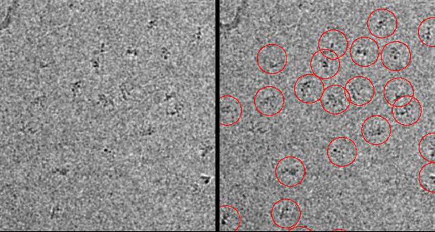
Using cryo-electron microscopy — which involves freezing, then imaging a sample — the researchers have taken thousands of photos of their current target, a protein complex involved in breaking down other, unwanted proteins. Each image contains 10 to 100 copies of the complex. It takes that many images to capture a protein from every angle. Once the 2-D images are stitched together, researchers can reconstruct the protein’s globular, 3-D shape at near-atomic resolution.
Microscopy Masters enlists volunteers to do the necessary first step of combing through the photos to find the protein molecules — a time-consuming job that people do better than computers. The task may feel daunting, as each black-and-white image resembles a fuzzy TV screen. Only some of the dark smudges in any given image will be molecules of interest; others will be actual smudges or globs of proteins too jumbled to be of use. Fortunately, a practice tutorial offers a crash course in protein identification. And each image will be classified by many users, alleviating some of the pressure of worrying about marking the wrong thing.
Data from the project will help researchers improve protein-picking computer algorithms, says project member Jacob Bruggemann. That way computers can take over the painstaking work.