New technique shows cells’ molecules in color
Electron microscopy is finally getting its Kodachrome moment.
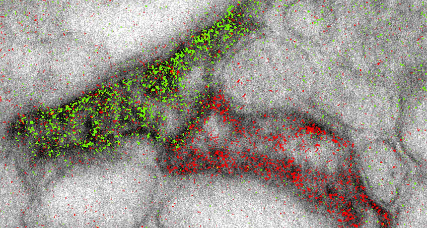
The high-powered scopes can now produce images that simultaneously highlight different molecules in different colors, scientists report online November 3 in Cell Chemical Biology. That’s helpful for researchers hoping to visualize the complex structures of cells or tissues — such as connections between brain cells, shown here.
Electron microscopes build black-and-white images by shooting beams of electrons at samples. Previously, scientists could add color by overlaying lower-resolution images from light microscopes. The new technique adds pizzazz without sacrificing image quality. It involves sequentially layering different metal ions on top of the sample. Each ion selectively latches onto a different target molecule. The electron beam interacts differently with each ion, yielding signature wave shapes that can be converted into colors. The researchers used the coloring technique to show that two brain cells called astrocytes (the edge of one shown in green, the other in red) could link up to the same message-sending junction between nerve cells.